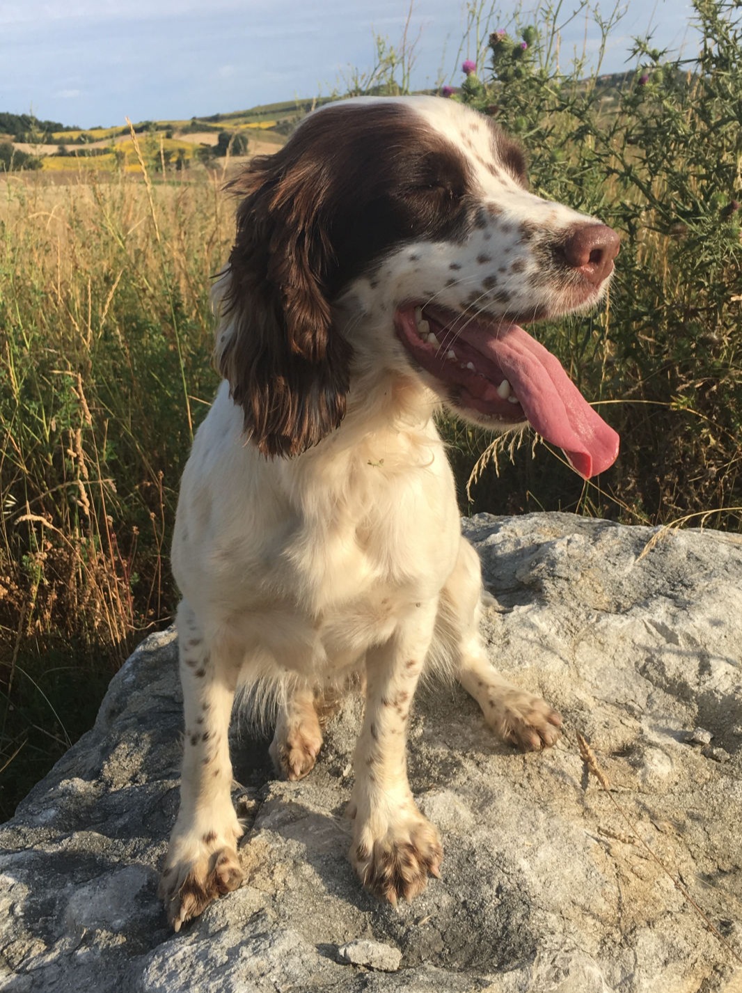
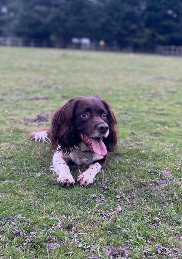
Bruce

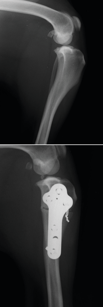
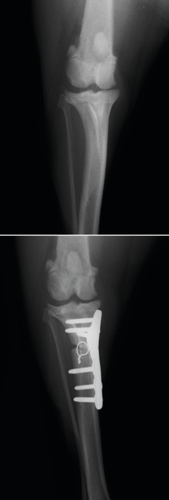
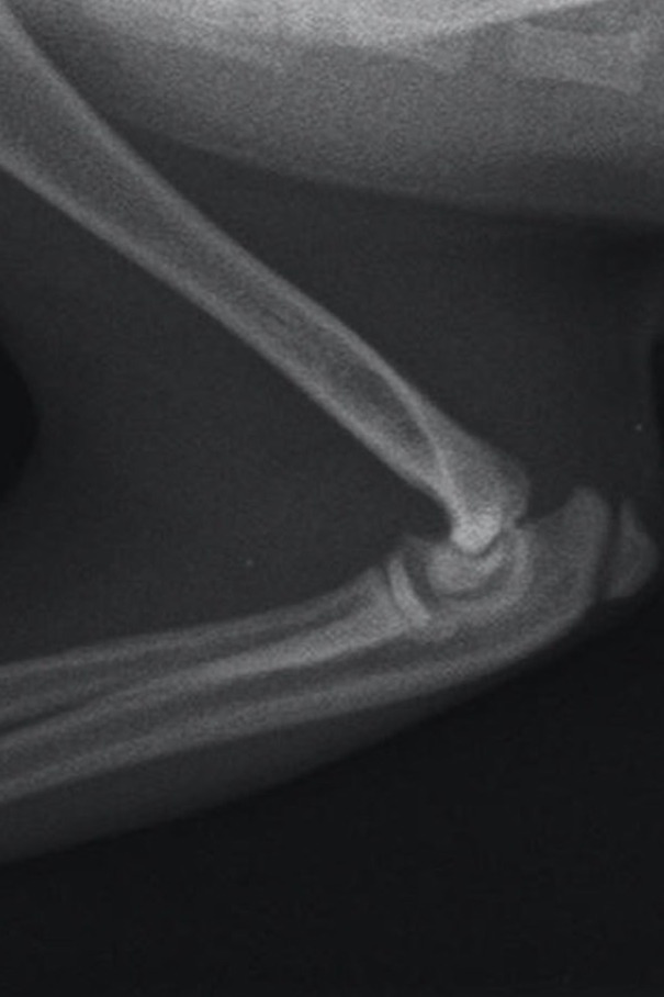
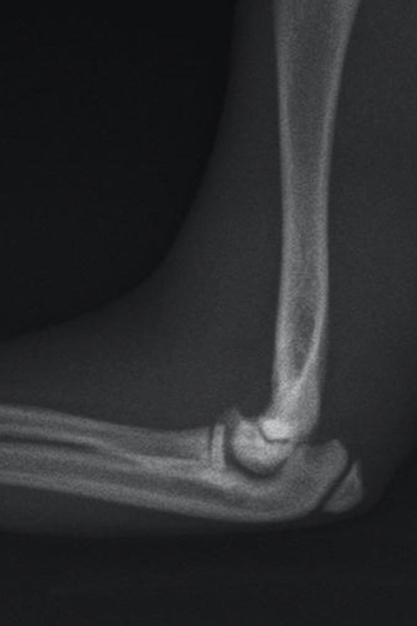
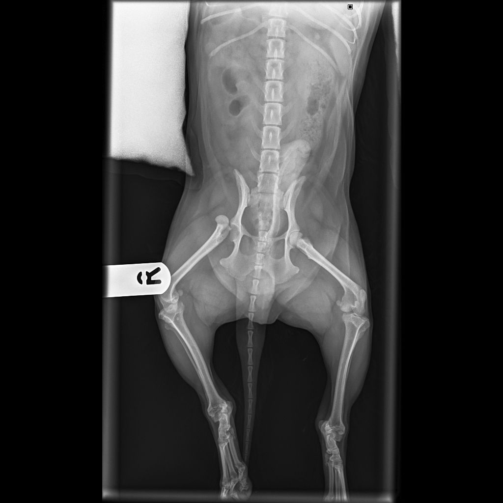
Bruce, the very handsome 9 year 8-month-old English Springer Spaniel, presented to his primary care practice in Spring 2023 having become acutely lame on his left pelvic limb. On examination, pain was elicited to his stifle so initial thoughts turned to potential cruciate disease however, as Bruce was otherwise well, his vet recommended a period of conservative management including restricted exercise and the introduction of a non-steroidal anti-inflammatory drug. Unfortunately, Bruce remained lame, so x-rays were the next step.

A free x-ray report was turned around within 24 hours and Bruce was booked in for a plateau levelling procedure; specifically a Cranial Closing Wedge Ostectomy (CCWO), the preferred technique of attending surgeon, Nicci.

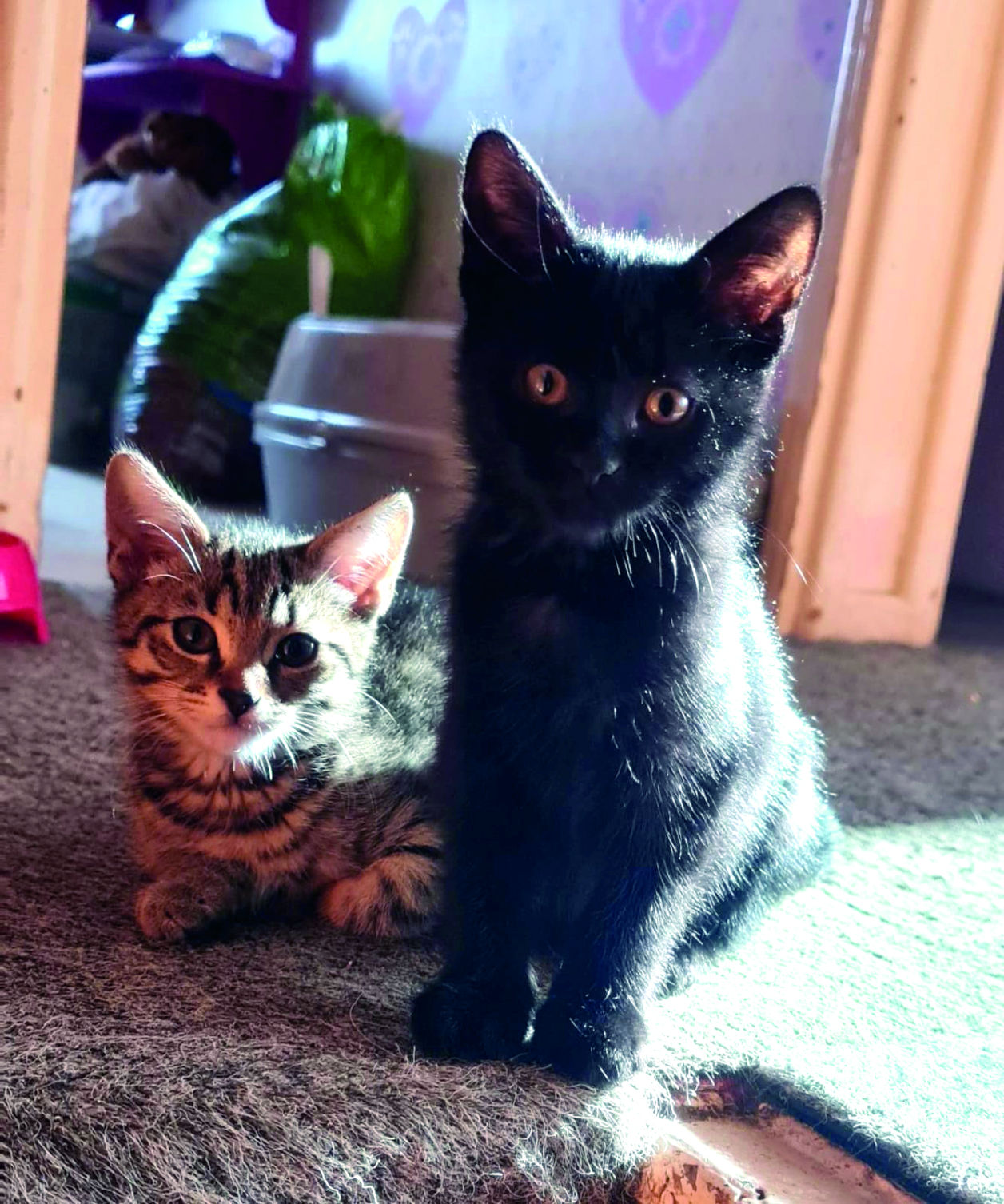
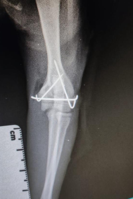
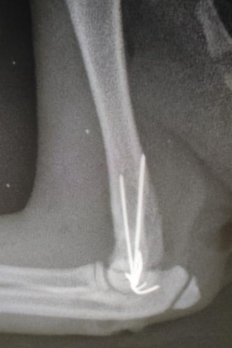
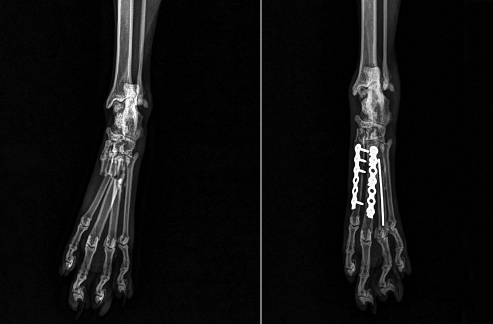
On Bruce’s surgery day, Nicci met with his family, discussed the procedure and answered all their questions before admitting him. A medial mini arthrotomy revealed complete rupture of the cranial cruciate ligament, while confirming that the menisci were intact. CCWO was performed with a 20-degree ostectomy, reduced with orthopaedic wire and stabilised with a 3.5mm N2 TPLO plate in compression. Post op x-rays revealed that implants were well placed so Bruce was recovered from his anaesthetic ready to begin his rehab programme. On Bruce’s return home, he was blessed with the care of his exceptionally doting family who made sure that he had all the home comforts he could possibly wish for – a king- size duvet and electric log burner were just a few mentioned! One particular family member that couldn’t see Bruce on his own was Margot, his feline – sister, pictured here looking over her ‘big - brother’s’ temporary castle. Short lead walks and controlled physio exercises were the key to Bruce’s journey before ‘X-ray day’ then came – offering great news as Nicci could confirm that Bruce’s osteotomy was healing nicely. Clinical exam also showed improved joint comfort, no lameness and an all-round happier Bruce, and family! Further rehab commenced after this time, giving Bruce the opportunity to put his ‘sea-legs’ on and take to the under-water-treadmill, hydrotherapy pool and wobble boards at Beacroft Referrals. Recent communication with Bruce’s family has been positive.

Radiographs of the limb (pre and post operative)

Radiographs of the limb (pre and post operative)

Boo

Boo is a 16-week-old kitten who had an unlucky fall whilst playing with his housemate at home and presented with an unusual elbow fracture involving the distal humeral physis. His fracture was repaired with tiny 0.6mm arthrodesis wires during a very delicate surgery to allow continued growth at the physis. Early stabilisation and perfect anatomical reduction was very important to allow a good functional outcome for this fracture. We are pleased to report that following 4 weeks of crate rest Boo has made an excellent recovery.

Physeal, or Salter Harris fractures are fractures that occur in skeletally immature patients and involve the growth plates or physes. These fractures can occur with minimal or no trauma, and patients can present following vigorous play, jumping or running. There are several different configurations of Salter Harris fractures. One of the most common that we see is a tibial crest avulsion fracture in young, well-muscled breeds such as French Bulldogs and Staffordshire Bull Terriers.

Flexed mediolateral radiograph

Neutral mediolateral radiograph

Post operative craniocaudal radiograph

Post operative mediolateral radiograph

Cadi

Cadi is a young working Springer Spaniel who's owners started to notice intermittent lameness affecting both the left and the right thoracic limbs. Plain radiographs were taken at Cadi's referring practice, but these images didn't reveal the cause of the lameness. Cadi was referred for a CT scan which showed she had bilateral Humeral Intracondylar Fissures (HIF), a condition most commonly seen in Spaniels, particularly Springer Spaniels.
The aetiology of this condition remains unclear, although it is thought that HIF lesions are stress fractures, rather than a developmental abnormality, as was previously thought. If left untreated, HIF lesions can predispose patients to fractures of the humeral condyle, or even complicated Y fractures of the distal humerus. Stabilisation of the HIF lesion with a transcondylar screw reduces lameness and the possibility of a fracture affecting this region.

From Cadi's CT scan, we were able to work with Fusion Implants www.fusionimplants.com to plan the optimal position of the transcondylar screws and prepare custom 3D printed drill guides to enable the screws to be placed accurately.

Cadi has made an excellent recovery post-operatively and is very much looking forward to returning to work in the next shooting season!

Radiographs of the right limb (pre and post operative)

Radiographs of the left limb (pre and post operative)

CT images planning the position of the 3D printed custom drill guide for placing the transcondylar screws.

Buzzy

Buzzy is a rescue yorkshire terrier, and one of our smaller canine patients at just over 3 Kg. Buzzy was involved in a dog fight which resulted in right coxo-femoral luxation. Buzzy's hip was reduced shortly after luxation, but as is often the case, Buzzy's hip reluxated and his owners opted for surgical stabilisation. Buzzy's hip was explored surgically and torn soft tissue and a haematoma was removed from the acetabulum. The coxofemoral joint was stabilised with a toggle system which involves passing a small metal rod (the toggle) attached to high tensile braided nylon through a drilled bone tunnel in the femoral neck, head and acetabulum before tensioning and securing the nylon against the femur. After a period of rest, Buzzy made an uneventful recovery from surgery.

Post-operative radiograph showing reduced coxofemoral joint and toggle repair

Pre-operative radiograph showing craniodorsal coxofemoral luxation

Kitty

Kitty is a 13 year old domestic short haired cat who found herself in trouble after falling from a work surface and fracturing three of her metatarsal bones including two of the main weight bearing metatarsals. The main challenge in repair of these fractures was the small diameter of the metatarsal bones, at only 4 mm across they would only accommodate the tiniest of implants.
A locking 1.5 kit was used to apply dorsal plates to metatarsals 2 and 3, and metatarsal 4 was repaired with a dowel pin due to the small amount of proximal bone stock.